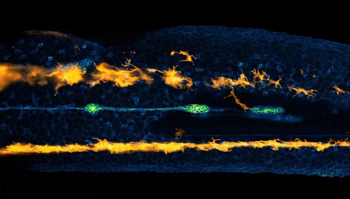
 Are you ready to peer inside the body of a fish embryo? Let's look into a small world! (Screenshot of Nikon Small World in Motion/Eduardo Zattara)
If there's one thing that is true about the world, it is that life looks different under a microscope.
Like, really different!
You could take the most seemingly everyday thing—a rock, a leaf, some dirt—and place it under a microscope, and you'd see some of the most wonderful and surprising details. And the more complex the object, the more spellbinding those details are.
This kind of beauty is what Nikon Small World is all about. Since 1975, this competition has combed through the year's best images taken with a microscope, bringing 'small worlds' some big attention.
In 2011, they expanded the competition to include video submissions, too. Called Small World in Motion, it has been always full of some amazing stuff. This year's winners have just been announced and we're so excited to share them with you right now.
Get ready to dive into these small worlds!
First place
The winner was a video is called 'Lateral line cells and melanocytes migrating in a zebrafish embryo.' The lateral line is a sensory organ found in many fish. So we're watching this organ in action in an unhatched fish baby. Cute!
It was taken by Dr. Eduardo Zattara of the National Scientific and Technical Research Council Bariloche, in Rio Negro, Argentina.
Second place
Next up is a 12-hour time-lapse of cells from a monkey. (A time-lapse is a video that shows a long stretch of time in a very short period of time.) The orange stuff is the membrane (outer layer) of the animal's plasma (blood), while the blue stuff is its DNA. (Great colour choice since DNA is an animal's blueprint, haha!)
This was made by Dr. Christophe Leterrier of the NeuroCyto Lab in Marseille, France.
Third place
And finally, in third place, is a really lovely look at the stinging cells of a sea anemone. The stringy lines across the video are the animal's neurons. This is what carries the electrical impulses that go from the brain to the rest of the body.
This video was made by Dr. Ahmet Karabulut of the Stowers Institute for Medical Research in Kansas City, Missouri, USA.